Custom Multiplex Imaging & Histology CRO Services
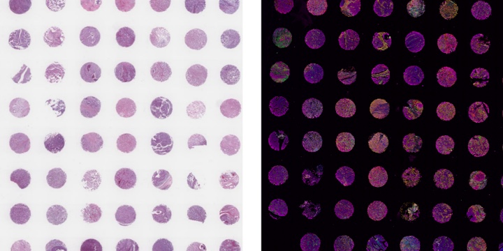
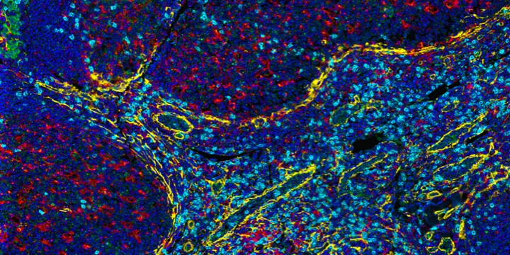
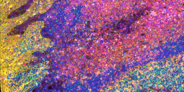
Multiplex immunofluorescence (mIF) is a powerful technique that allows researchers to simultaneously visualize and analyze multiple components of the tissue microenvironment. Multiplexing uses multiple antibodies with fluorescent detection to target specific proteins within the microenvironment, allowing researchers to create a detailed map of the various cells, proteins and their interactions. We offer both low plex and high plex options for mIF which allow you to profile tissues to determine cell phenotypes, their functional state, and cell interactions.
Key Service Offerings
- Core panels encompassing basic human I-O phenotypic markers
- Up to 40-plex discovery panels
- Custom validation of antibodies of your choosing
- Library of validated antibodies to choose from
- Creation of custom panels
- Antibodies can be combined with a core panel to create higher plex panels
- Histology services to prepare tissue for staining

We can support your needs across the entire imaging workflow
Our Equipment
PhenoImager™ HT
Services
- Up to 6-plex panel using Opal™ reagents
- Panel optimization and antibody order testing
- Library of validated antibodies
- Sample staining
- Slide scanning
Specs
- 0.25 microns/pixel
- Separate 7-colors (DAPI plus 6 antibodies)
- Capable of large tissue microarrays
- Low plex, high throughput, 80 slide capacity
Lunaphore COMET™
Services
- Runs of 40-plex using seqIF™
- Core Panels available, including SPYRE™ Antibody Panels from Lunaphore
- Addition of other catalog or custom antibodies to Core Panel
- Creation of bespoke panels
- Library of validated antibodies to choose from
Specs
- 0.23 microns/pixel
- 9×9 mm scan area can accommodate tissue sections and small tissue arrays
- No need for conjugated primary antibodies
- High plex, medium throughput
- 24 slides/week for 10-plex
- 20 slides/week for 20-plex
- 8 slides/week for 40-plex
Modular Panel Design
10× Core Panel for human I-O markers
Marker |
Catalog # |
Host Species |
Relevance |
|---|---|---|---|
CD3 |
Rb |
T-cells |
|
CD20 |
Mo |
B-cells |
|
FoxP3 |
Rb |
Regulatory T-cells |
|
CD8 |
Mo |
Cytotoxic T-cells |
|
PD-L1 |
Rb |
Checkpoint |
|
Pan-CK |
Mo |
Epithelium |
|
PD-1 |
Rb |
T-cells |
|
CD68 |
Mo |
Macrophages |
|
CD45 |
Rb |
Lymphocytes |
|
PCNA |
Mo |
Proliferation |
EMT Panel
Target |
Clone |
Relevance |
|---|---|---|
Beta-catenin |
BLR086G |
Transcription factor |
Vimentin |
BLR100G |
Mesenchymal |
ZEB1 |
BLR217K |
Transcription factor |
EpCAM |
BLR077G |
Epithelial |
E-cadherin |
BLR088G |
Epithelial |
SOX10 |
BLR080G |
Mesenchymal |
ZO-1/TJP1 |
BLR092G |
Epithelial |
ASMA |
BLR082G |
Epithelial |
T-cell Profiling Panel
Target |
Clone |
Relevance |
|---|---|---|
GATA3 |
BLR121H |
Th2 naive T cell |
T-bet |
BLRI10H |
Th1 naive T cell |
CD25 |
BLR158J |
Regulatory T cell; Activated |
CD4 |
BL-155-1C11 |
Helper cells |
CD45RO |
UCHL-1 |
Memory T cells |
Granzyme B |
BLRO22E |
Cytotoxic T cell |
Breast Cancer Module
Target |
Clone |
Relevance |
|---|---|---|
EGFR |
BLR252L |
tumorigenesis |
PR |
188 |
modulates ERa; prognostic marker |
HER2 |
BLR241L |
oncogene; prognostic marker |
ERa |
119-13 |
prognostic marker; linked with PR |
Cytokeratin (Structural) Module
Target |
Clone |
Relevance |
|---|---|---|
CK7 |
BC1 |
Type II, nonkeratinizing epithelia |
CK14 |
LLO02 |
Type I, stratified epithelium |
CK18 |
LDK18 |
Type I, simple epithelia |
CK19 |
BA17 |
Simple and complex epithelium |
СК20 |
BLR105H |
Type I, mature enterocytes |
- Extensive collection of in-house manufactured antibodies to choose from to customize your assay
- Immune cell markers
- Cytokeratin markers
- Cancer markers
- Phospho-proteins
- Signaling pathway
- Neurobiology markers
- Mouse proteins
- Assistance with sourcing other antibodies
Full Service Histology Lab
- Sectioning
- Tissue microarray
- H&E staining
- Whole Slide Scanning
Our Partners


Additional Resources
- Immunofluorescence
- Multiplex mIHC: Making Discoveries in Multicolor
- Tyramide Signal Amplification (TSA)-Based Immunofluorescent Multiplex (mIF) Assays
- Antibody validation for successful immunohistochemistry and multiplex immunofluorescence
- Focus on Immuno-oncology and Immunotherapy
Custom Request
Fortis Life Sciences is a registered trademark of Fortis Life Sciences, LLC.
COMET, seqIF, and SPYRE are registered trademarks of Lunaphore Technologies S.A.
PhenoImager, Opal, and Akoya Biosciences are registered trademarks of Akoya Biosciences, Inc.
By clicking “Acknowledge”, you consent to our website's use of cookies to give you the most relevant experience by remembering your preferences and to analyze our website traffic.


