Focus on Autophagy
The Focus on Autophagy is a collation of articles about this important cellular process. Learn about autophagy and the products that Fortis offers to study this pathway, and sign up to receive emails about future Focus on… features.

Inside the cell, there are multiple mechanisms to degrade intra- and extra-cellular components. Autophagy is a non-specific pathway that sequesters cytoplasmic contents via double membranes to recycle intracellular contents. It was initially characterized as a starvation response. When cells are depleted of nutrients, they must break down cellular components that are not necessary to supply the building blocks for those that are. This recycling process – autophagy – is more efficient than creating or manufacturing individual macromolecules. Autophagy is critical for cell and tissue homeostasis and when disrupted, can lead to a variety of disease states including cancer, increased pathogen replication, heart disease, pro-aging, and neurological disorders.
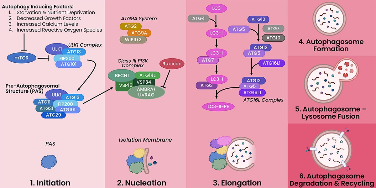
The process of autophagy can be broken down into six steps.
- Initiation: the clustering of autophagy factors to create the core autophagy complex
- Nucleation: the formation of the initial double membrane at the site of the core autophagy complex
- Elongation: the extension of the double membrane
- Autophagosome Formation: the complete enclosure of cytoplasmic contents by the double membrane forming the autophagosome
- Autophagosome-Lysosome Fusion: the fusion of the lysosome and the autophagosome, exposing the contents of the autophagosome to the degrading enzymes of the lysosome
- Degradation & Recycling: the breakdown of the autophagosome contents by lysosomal enzymes resulting in building blocks that can be released back into the cytoplasm and used to make DNA, RNA, proteins, lipids, and carbohydrates

Cutting-edge Autophagy Research
USP11 promotes autophagy
USP11 promotes autophagy by directly stabilizing Beclin-1. A recent publication identifies USP11 as a potential therapeutic target to inhibit autophagy in cancer.
CD38 and LRRK2 play a role in autophagy induction
A recent paper by Neel Nabar and colleagues from the National Institute of Health added new signaling molecules (CD38 and LRRK2) into the autophagy induction pathways.
Generation and Application of a Novel Monoclonal Antibody to UBQLN4 to Characterize the dsDNA Damage Response
Ubqln4 plays an important role in autophagy by interacting with the autophagosome membrane and recruiting additional proteins required for autophagosome-lysosome fusion. Learn how Fortis made a highly specific antibody for Ubqln4, using our six pillars of validation approach, that can be used in WB, IP, flow cytometry, and multiplex IHC.
KANSL1 is a regulator of autophagosome-lysosome fusion
A recent study shows a link between autophagy and a rare genetic disorder, Koolen-de Vries syndrome, caused by loss of KANSL1. An FDA-approved drug, 13-cis retinoic acid, was able to rescue autophagy in the absence of KANSL1.
FKBP51 links autophagy and obesity
FKBP51 expression levels tightly control induction or inhibition of autophagy. In the mediobasal hypothalamus, this regulation of autophagy has a profound impact on obesity.
MondoA controls senescence through autophagy and mitochondrial homesostasis
A recent study provides mechanistic insights for the involvement of autophagy in cellular senescence. This research shows the transcription factor MondoA controls expression of the autophagy inhibitor Rubicon when senescence is induced.
Sign up to receive future Focus on… features
Fortis Products for Studying Autophagy
Don’t see the see the target you are looking for? Let us know.
|
Name |
Role in Autophagy |
Catalog # |
Applications |
Reactivity |
Host |
Clonality |
|
AMBRA1 |
Positive regulator of autophagy |
IP, WB |
Hu |
Rabbit |
Polyclonal |
|
|
AMPK |
Autophagosome maturation & lysosome fusion |
IHC, IP, WB |
Hu, Ms |
Rabbit |
Polyclonal |
|
|
AMPK |
Autophagosome maturation & lysosome fusion |
IP, WB |
Hu, Ms |
Rabbit |
Polyclonal |
|
|
ATG4B |
Processing and recycling of LC3. Facilitates autophagosome-lysosome fusion. |
IP |
Hu |
Rabbit |
Polyclonal |
|
|
ATG4B |
Processing and recycling of LC3. Facilitates autophagosome-lysosome fusion. |
IP, WB |
Hu, Ms |
Rabbit |
Polyclonal |
|
|
ATG9A |
Interacts with PAS and isolation membrane formation. Generation of phagophore. |
IP, WB |
Hu |
Rabbit |
Polyclonal |
|
|
ATG13 |
Initiates autophagy |
IP, WB |
Hu, Ms |
Rabbit |
Polyclonal |
|
|
ATG16L1 |
Determines site of LC3 conjugation. Facilitates membrane elongation. |
IP, WB |
Hu |
Rabbit |
Polyclonal |
|
|
ATG16L1 |
Determines site of LC3 conjugation. Facilitates membrane elongation. |
IP |
Hu |
Rabbit |
Polyclonal |
|
|
Beclin-1 |
Autophagic protein localization to PAS |
IP, WB |
Hu, Ms |
Rabbit |
Polyclonal |
|
|
Beclin-1 |
Autophagic protein localization to PAS |
IP |
Hu |
Rabbit |
Polyclonal |
|
|
EI24 |
Formation of autophagosome-lysosome fusion and degradation of contents. |
IP |
Hu |
Rabbit |
Polyclonal |
|
|
FIP200 |
Stability and phosphorylation of ULK1 resulting in initiation of autophagy. |
IP, WB |
Hu, Ms |
Rabbit |
Polyclonal |
|
|
FIP200 |
Stability and phosphorylation of ULK1 resulting in initiation of autophagy. |
IP |
Hu |
Rabbit |
Polyclonal |
|
|
FYCO1 |
Binds LC3 to facilitate autophagosome-lysosome fusion. |
IP, WB |
Hu |
Rabbit |
Polyclonal |
|
|
FYCO1 |
Binds LC3 to facilitate autophagosome-lysosome fusion. |
IP, WB |
Hu |
Rabbit |
Polyclonal |
|
|
mTOR |
Inhibits autophagy |
ICC, IP, WB |
Hu |
Rabbit |
Polyclonal |
|
|
mTOR |
Inhibits autophagy |
IP, WB |
Hu |
Rabbit |
Polyclonal |
|
|
mTOR |
Inhibits autophagy |
IP, WB |
Hu, Ms |
Rabbit |
Polyclonal |
|
|
mTOR |
Inhibits autophagy |
IP, WB |
Hu |
Rabbit |
Polyclonal |
|
|
MondoA |
Activates autophagy |
IP, WB |
Hu |
Rabbit |
Polyclonal |
|
|
P62/ |
Guides polyubiquitinated proteins & organelles to autophagosomes |
IHC, IP, WB |
Hu, Ms |
Rabbit |
Polyclonal |
|
|
P62/ |
Guides polyubiquitinated proteins & organelles to autophagosomes |
IHC, IP, WB |
Hu |
Rabbit |
Polyclonal |
|
|
P62/ |
Guides polyubiquitinated proteins & organelles to autophagosomes |
IHC, IP, WB |
Hu, Ms |
Rabbit |
Polyclonal |
|
|
Rubicon |
Inhibits Class III PI3K Complex via interaction with UVRAG. |
IP, WB |
Hu |
Rabbit |
Polyclonal |
|
|
SOGA1 |
Regulates autophagy |
IP |
Hu |
Rabbit |
Polyclonal |
|
|
TFEB |
Master regulator of autophagy |
IP, WB |
Hu, Ms |
Rabbit |
Monoclonal |
|
|
TFEB |
Master regulator of autophagy |
IP, WB |
Hu |
Rabbit |
Polyclonal |
|
|
TFEB |
Master regulator of autophagy |
IP, WB |
Hu, Ms |
Rabbit |
Polyclonal |
|
|
USP11 |
Promotes autophagy |
IP, WB |
Hu |
Rabbit |
Polyclonal |
|
|
UVRAG |
Promotes autophagy |
IHC, IP, WB |
Hu |
Rabbit |
Polyclonal |
|
|
VMA21 |
Decreases lysosomal pH enhancing autophagic degradation |
IP, WB |
Hu, Ms |
Rabbit |
Polyclonal |
|
|
VMA21 |
Decreases lysosomal pH enhancing autophagic degradation |
IP, WB |
Hu, Ms |
Rabbit |
Polyclonal |
|
|
VPS15 |
Promotes autophagy |
IP |
Hu |
Rabbit |
Polyclonal |
|
|
VPS15 |
Promotes autophagy |
IP, WB |
Hu, Ms |
Rabbit |
Polyclonal |
|
|
WDFY3 |
Associated with autophagic membranes |
IHC, IP, WB |
Hu |
Rabbit |
Polyclonal |
By clicking “Acknowledge”, you consent to our website's use of cookies to give you the most relevant experience by remembering your preferences and to analyze our website traffic.
